|
Lymphedema,
a protein-rich swelling which usually affects the
extremities, is a very common condition worldwide. Complete
Decongestive Physiotherapy is done successfully
in Europe for decades in the treatment of primary and
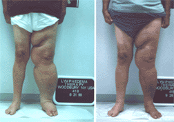
secondary Lymphedema. Chronic peripheral lymphedema, an accumulation of protein-rich fluid in the superficial tissues, is a very common and serious condition with significant consequences for the patient. One of the main reasons for the development of lymphedema are surgical interventions in combination with lymph node dissections, such as mastectomy or lumpectomy due to breast cancer. Although reliable statistics on the overall incidence of lymphedema are not available, conservative numbers estimate the incidence of secondary upper extremity lymphedema to be around 1 per cent of the population. In addition to that there is a large number of patients suffering from primary lymphedema, which usually affects the lower extremities and is caused by congenital malformations of the lymphatic system. Since lymphedema, primary or secondary, is a progressive condition, treatment should begin as early as possible. The goal of the treatment is to remove the excess lymphatic loads of water and protein and to restore the disturbed equilibrium in the interstitial tissues of the affected area. Treatment of Lymphedema with Complete Decongestive PhysiotherapyComplete Decongestive Physiotherapy (CDP), done successfully in Europe for decades, is a non-invasive therapy with long lasting results. CDP is superior to all other approaches to treat lymphedema (pumps, medication, surgery) and designed to reduce and to maintain the reduction of the swollen extremity. At the end of the 19th century, Alexander von Winiwarter, Professor of Surgery, already described the basic steps of this therapy of swollen limbs with special massage, compression, and elevation. The technique was improved in the 1930s by Vodder, a physical therapist from Denmark, who successfully treated lymphedema. In the 1980s Prof. Michael Foeldi, M.D. considerably improved this therapy by developing a technique called Complete Decongestive Physiotherapy, which even in advanced stages of lymphedema shows remarkable results. CDP is done in two phases. The first phase, intensive phase, lasts between two and four weeks (in extreme cases longer), treatments are done twice a day, five days a week. The goal of this phase is to decongest the swollen extremity to a normal or near normal size. Simultaneously the patient is instructed in techniques designed to maintain and even improve the condition after the intensive phase of the therapy. The first phase is immediately followed by phase two, the maintenance and improvement phase which the patients continues at home. For the safety of the patients and to achieve good results it is absolutely mandatory that the therapist is thoroughly trained in all components of CDP. Only certified MLD/ CDP therapists have a complete understanding of the pathophysiology of lymphedema and its treatment. CDP consists of four basic steps:
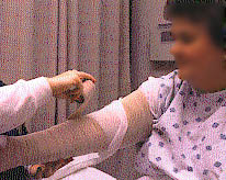
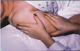
Manual Lmphatic Drainage (MLD) is a gentle manual treatment technique which improves the activity of intact lymph vessels by mild mechanical stimuli, the goal of this treatment is to move excess lymphatic loads of water and protein into areas with still sufficient lymphatics. This is a very light special skin technique which is directed toward stimulating the activity of the lymphangions, designed to empty and decompress the obstructed lymph vessels. MLD allows the natural pattern of existing vessels located under the skin and areas where the lymph system and venous system join to assume the function of transporting lymph fluid. The hands of the therapist are used to soften fibrotic lakes which can accumulate under the skin. The areas located away from the limb are always treated first. MLD techniques are very specific and involve pressures and release of pressures along with strokes. Since the elastic fibers of the skin are destroyed during the course of lymphedema it is mandatory to apply sufficient compression to the affected area in order to prevent re-accumulation of fluid. Compression in phase one is achieved by short-stretch bandages applied after each treatment. It consists of 4 layers of material that protect lymphatics, offer gradient compression, and soften the skin. Bandaging does not permit the treatment areas to refill with fluid. After the extremity is decongested the patient if fitted with a sufficient compression garment that needs to be worn during the daytime. At night the patient applies mild compression using bandages. Before treatment can be started, the skin has to be free of infections or fungal affections. During treatment a low-pH lotion is applied to maintain the moisture of the skin. Remedial exercises performed by the patient wearing the compression bandage or garment aid the lymphokinetic effects of the joint and muscle pumps. Muscle activation with the bandages in place provides resistance. When resistance is met, increased lymph flow and reduction is the result. The bandaging is bulky and will not accommodate tight fitting clothing:
Anatomy and Physiology of the Lymphatic SystemUnlike the blood system the lymphatic system works according to the one way principle, its main purpose is to transport "waste materials" from the interstitial tissues back into the blood system. These materials, also called lymphatic loads, consist of protein, water, cells and fat, are drained by the various vascular structures of the lymphatic system and filtered by a large number of regional and central lymph nodes before they enter the venous system. Part of these waste materials are also cell products and cell residues including foreign materials. Initial lymphatic system: Lymph vessels start in almost every tissue as lymph capillaries. These initial lymphatics are made up of endothelial cells which overlap each other. Lymph capillaries do not have a continuous connection like blood capillary endothelial cells do. A surrounding fibre net, anchoring filaments, arranged around the lymph capillaries, enables these small vessels to stay open, even under high tissue pressure. Lymph capillaries collect lymphatic loads from the interstitial areas and gradually join together into bigger lymph vessels, so-called precollectors which then drain into collectors. Collectors: One segment of a lymph collector is called lymph angion. Contractions of smooth muscles situated in each lymph angion, generate the propulsive force of the lymph flow. The pumping is aided by a large number of valves located inside the collectors which allow the lymph flow in only one direction. After passing a large number of lymph nodes, where foreign substances like bacteria, are filtered out and necessary immune reactions are activated, the lymph fluid empties into the venous system, mainly via the thoracic duct. The thoracic duct is the largest lymph vessel of the body. Under physiological conditions approximately 1-2 liters of lymph fluid drain in 24 hours via the thoracic duct into the left venous angle, formed by the left internal jugular and the left subclavian vein. Starling's equilibrium: The amount of water and protein transported via the lymphatic system is depending on forces being active in the area of the blood capillaries. Starling's equilibrium describes the balance of capillary filtration and capillary reabsorption. The transport of fluid through the membrane of blood capillaries depends on four variables: 1. blood capillary
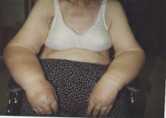
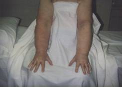
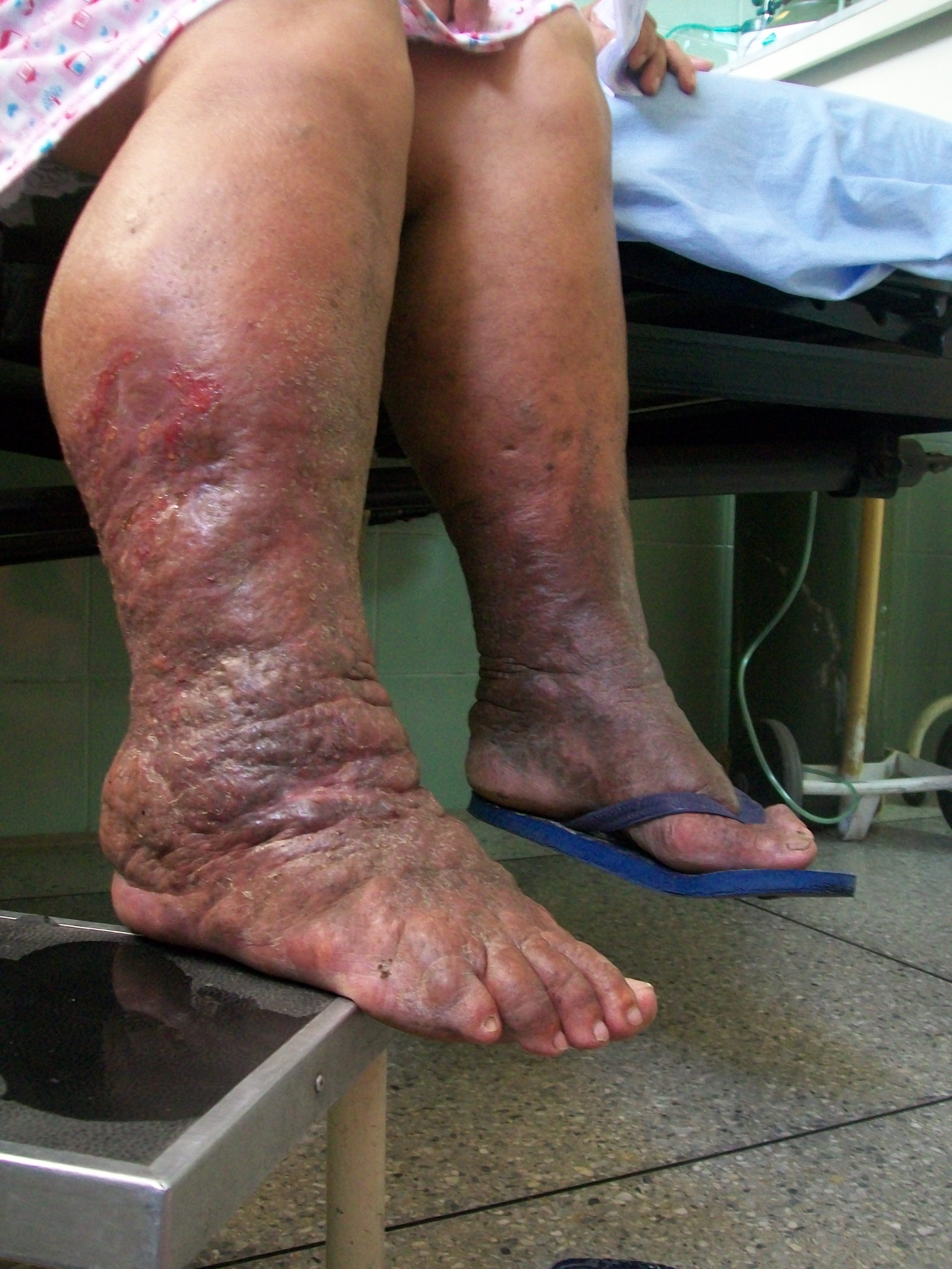
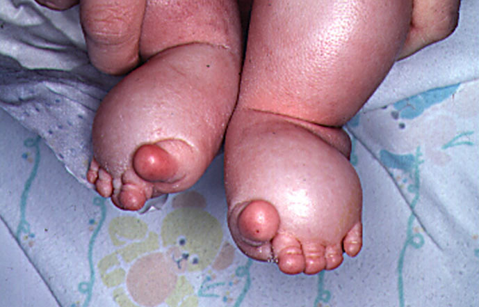
pressure (BCP) Ultrafiltration: Reabsorption: Under physiological conditions 10-15% of the ultrafiltrate remains in the interstitial tissues and is then drained by the lymphatic system. Shifting of Starling's equilibrium towards an increase in ultrafiltration, e.g. increased blood capillary pressure (inflammation, venous hypertension) or decreased colloidosmotic pressure (hypoproteinemia), can cause an increased amount of water and proteins, thus creating a higher burden on the lymphatic system. A healthy lymphatic system is, for some time, able to prevent the onset of edema, under normal conditions the transport capacity (TC) of the lymphatic system is approximately 10 times higher than the physiological amount of the lymphatic loads (LL) of water and protein = functional reserve (FR) of the lymphatic system. Pathophysiology of Lymphedema:As long as the lymphatic loads remains lower than the transport capacity of the lymphatic system, the lymphatic compensation is successful. If the amount of water and protein exceeds the transport capacity, edema will occur. This condition is called dynamic insufficiency of the lymphatic system, the lymph vessels are intact but overwhelmed. The result is an accumulation of fluid in the tissue which is usually treated successfully with elevation of the affected limbs, compression and decongestive exercises. Lymphedema is caused by a mechanical insufficiency, or low-volume insufficiency of the lymphatic system. In this case the transport capacity of the lymphatic system drops below the physiological level of the lymphatic loads of water and protein, that means the lymphatic system is not able to manage its main purpose which is to clear the interstitial spaces from excess water, protein and other chemical, organic and inorganic cell products. The reasons for mechanical insufficiencies are various and mainly caused by surgery, radiation, trauma or inflammation. Accumulation of high protein fluid is the result which is then recognized as lymphedema or lymphostatic edema. Any lymphedema left untreated gradually worsens and will have significant consequences.The first stage of lymphedema (reversible stage) is characterized by a smooth texture of the tissue, the lymphedema is "pitting" and may vanish more or less over night. If protein rich edema persists, the congested protein molecules are replaced by fibrotic tissue, i.e. the lymphedema becomes harder (stage II) and in addition to that patients are prone to developing frequent infections which additionally worsen the condition. Typical for the third stage of lymphedema, lymphostatic elephantiasis, is an extreme increase of the swelling combined with skin changes, loss of function and other complications. Since lymphedema, primary or secondary, is a progressive condition, treatment should begin as early as possible. The goal of the treatment is to remove the excess lymphatic loads of water and protein and to restore the disturbed equilibrium in the interstitial tissues of the affected area. |
|
|
||
| Homepage |